How do you do a wet mount on a microscope?
How do you do a wet mount on a microscope?
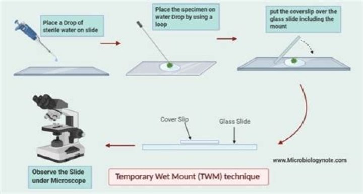
Wet-mount Slides
- Collect a thin slice of your sample and place it on a clean, dry slide.
- Place one drop of water over your sample.
- Place the coverslip at a 45-degree angle with one edge touching the water and let go.
- Your slide is ready to be viewed.
How do you prepare cheek cells for viewing under the microscope?
Methods
- Take a clean cotton swab and gently scrape the inside of your mouth.
- Smear the cotton swab on the centre of the microscope slide for 2 to 3 seconds.
- Add a drop of methylene blue solution and place a coverslip on top.
- Remove any excess solution by allowing a paper towel to touch one side of the coverslip.
When preparing a wet mount like the cheek cells what object should be carefully laid over the specimen?
Carefully lower a cover slip over the stain/cheek scrapings mixture. Note: Slowly lowering the cover slip at about a 450 degree angle helps avoid air bubbles getting trapped beneath the cover slip. This is called a temporary, wet mount, since a cover slip was placed over a specimen within a liquid.
How do you make a wet mount cheek epithelial cell?
How to Prepare a Wet Mount of Cheek Cells
- place a drop of physiological saline on a clean microscopic slide (central part of the slide)
- smear the cotton swab on to the center (part containing the saline drop) of the clean slide for about 4 seconds to get the cells on to the center of the slide.
How do you prepare a wet mount of cheek cells?
When preparing a wet mount for a study under the microscope What will you do to ensure that the liquid portion of the specimen will not spill on it?
Question: When preparing a wet mount for study under the microscope, you must ensure that a cover slip is placed over the specimen a drop of water is added to the specimen on the slide the low power lens is above the object on the stage a drop of methylene blue is added to the specimen.