How do you interpret IVUS?
How do you interpret IVUS?
The interpretation of IVUS relies on simple visual inspection of acoustic reflections to determine plaque composition. However, different tissue components may look quite similar, and artifacts may adversely affect ultrasound images. IVUS commonly detects occult disease in angiographically “normal” sites.
What is coronary IVUS?
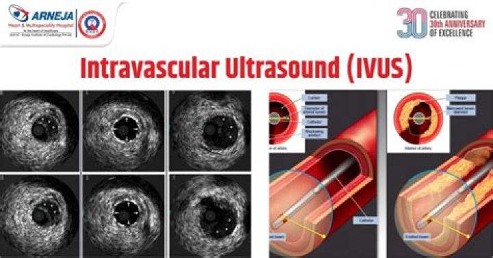
Intravascular Ultrasound (IVUS) is a catheter-based diagnostic procedure used to view the inside of a coronary artery, providing a real-time view.
What is MLA in IVUS?
Although the IVUS minimum lumen area (MLA) was the parameter that best correlated with ischemia, reported thresholds of IVUS MLA cut-offs ranged from 2.1 to 4.4 mm2, the thresholds were smaller in Asian studies than in Western studies, and the “most common” cut-off was approximately 3.0 mm2.
What does FFR measure?
Fractional flow reserve (FFR) measurement involves determining the ratio between the maximum achievable blood flow in a diseased coronary artery and the theoretical maximum flow in a normal coronary artery.
How do you use IVUS?
To use IVUS, physicians use a guide wire, usually 0.36 mm, and the IVUS-tipped catheter is then fed over the guide wire. Angiography is used to guide the IVUS catheter to the area of the vessel to be imaged. It is placed farthest away from the area to be imaged and is then pulled back through the area of stenosis.
How do you size a IVUS stent?
The traditional IVUS method requires measurement of the maximum proximal and distal reference lumen diameters by IVUS. The stent size is selected based on the larger of these measurements. The aggressive IVUS method requires measurement of the media-to-media dimensions in an orthogonal manner within a coronary lesion.
What is a negative FFR?
For the most part, a negative FFR predicts the ability to safely defer interventional treatment of an intermediate lesion, with a relatively low risk of downstream events.
How FFR test is done?
FFR uses a small sensor on the tip of the wire (commonly a transducer) to measure pressure, temperature and flow to determine the exact severity of the lesion. This is done during maximal blood flow (hyperemia), which can be induced by injecting products such as adenosine or papaverine.
What does PTCA mean in medical terms?
PTCA, or percutaneous transluminal coronary angioplasty, is a minimally invasive procedure that opens blocked coronary arteries to improve blood flow to the heart muscle.
Does IVUS use contrast?
Conclusions. Thoughtful and extensive use of IVUS as the primary imaging tool to guide PCI is safe and markedly reduces the volume of iodine contrast used compared with guidance by angiography alone. IVUS imaging should be considered for patients at high risk of CI-AKI or volume overload undergoing coronary angioplasty …
What is OCT in coronary angiography?
Optical coherence tomography (OCT) is an optical analog of intravascular ultrasound (IVUS) that can be used to examine the coronary arteries and has 10-fold higher resolution than IVUS.