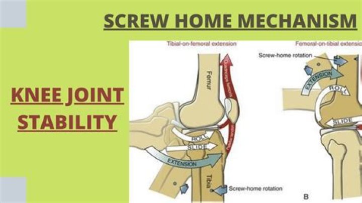
What features help Stabilise the knee joint
The lateral collateral ligament (LCL) stabilises the knee on the outside of the joint. Two crescent shaped bands of cartilage called the medial and lateral menisci act as shock absorbers between the femur and tibia. They protect the ends of the bones from rubbing on each other and help stabilise the knee further.
What structures help stabilize knee joint?
The ligaments of the knee function to stabilize the knee joint. There are two important groups of ligaments that hold the bones of the knee joint together, collateral ligaments and the cruciate ligament. Collateral ligaments are present on either side of the knee.
Which structures contribute to stabilizing the knee joint quizlet?
- crural fascia,
- the tibia, the.
- interosseous membrane,
- the fibula and the.
- anterior intermuscular septum.
What makes the knee joint stable?
Naturally the ilio-tibial band, the lateral collateral ligament, the popliteus tendon, the biceps tendon, the postero-lateral capsule and the lateral head of gastrocnemius are all important factors contributing to stability.How do you stabilize joints?
- Exercise Regularly. Exercise improves bone density and keeps the muscles that surround your joints strong, says A. …
- Build Muscle Strength. …
- Strengthen Your Core. …
- Try Low-Impact Cardio. …
- Stretch After Your Workout. …
- Prevent Exercise-Related Injury. …
- Lose Extra Weight.
What is responsible for maintaining the position of the patella during knee movement?
The patellar facets are convex in shape in order to accommodate the concave femoral surface with the lateral side wider to help maintain patellar position. The majority of the articulating surface of the patella is covered with a thick layer of articular cartilage, up to seven millimeters.
Which is the most important muscle which helps to stabilize the knee joint?
The two main muscle groups of the knee knee joint are the quadriceps and the hamstrings. Both play a vital role, both moving and stabilizing the knee joint. The quadriceps muscle group is made up of four different individual muscles which join together forming the quadriceps tendon.
What stabilizes the posterior knee joint quizlet?
The ACL stabilizes the knee joint by stretching diagonally from the femur at the back of the joint to the tibia in the front, which normally prevents forward or anterior movement of the tibia from underneath the femur. It also resists medial rotation of the tibia.What movement does the knee joint allow?
The knee joint is a modified hinge joint (ginglymus). The active movements of the knee joint are described as flexion, extension, medial rotation and lateral rotation.
What features keep the elbow relatively stable?The structures that stabilize the elbow include the coronoid process, the radial (lateral) collateral ligament, and the anterior portion of the ulnar (medial) collateral ligament.
Article first time published onWhat stabilizes the knee on the posterior side?
The muscles surrounding the knee function to both move and stabilize the joint. The two main muscle groups are the quadriceps on the anterior side of the knee and femur, and the hamstrings on the posterior side.
What 4 factors determine the stability of joints?
- Shape of articular surfaces (how close they fit)
- Strength and tension of capsule and ligaments (dependent on position)
- Arrangement and tension of muscles.
- Contact with soft parts such as adipose tissue.
- Hormones.
Which muscles help stabilize joint activity?
Skeletal muscles maintain posture, stabilize bones and joints, control internal movement, and generate heat. Skeletal muscle fibers are long, multinucleated cells.
How can physical activity improve joint stability?
“Exercise strengthens the muscles, ligaments and tendons surrounding the joints,” says Sterling. “When these tissues are strong, they act like a brace to protect the joint,” and lessen pressure on weakened joints.
How can I improve my knee strength and stability?
- Benefits.
- Leg lifts.
- Standing hamstring curls.
- Hamstring curls on a weight bench.
- Step exercises.
- Single-leg dip.
- Wall squats.
- Post-exercise stretching.
What ligaments stabilize the knee?
- Anterior cruciate ligament (ACL). …
- Posterior cruciate ligament (PCL). …
- Medial collateral ligament (MCL). …
- Lateral collateral ligament (LCL).
What stabilizes the patellofemoral joint?
Ligaments. The patellar retinaculum is an important stabilizer of the patellofemoral joint, mainly its medial and lateral components. The Medial Patellofemoral Ligament (MPFL) – originates on the medial femur and has a “sail-shaped” attachment on the patella and quadriceps tendon.
What ligaments stabilize the patella?
The medial patellofemoral ligament is a part of the complex network of soft tissues that stabilize the knee. The MPFL attaches the inside part of the patella (kneecap) to the long bone of the thigh, also called the femur.
Why is the knee joint so important in movement?
The knee joint is one of the strongest and most important joints in the human body. It allows the lower leg to move relative to the thigh while supporting the body’s weight. Movements at the knee joint are essential to many everyday activities, including walking, running, sitting and standing.
What reduces friction at joints?
Cartilage helps reduce the friction of movement within a joint. Synovial membrane. A tissue called the synovial membrane lines the joint and seals it into a joint capsule. The synovial membrane secretes a clear, sticky fluid (synovial fluid) around the joint to lubricate it.
What is the antagonist muscle in knee flexion?
Movement = starts off with knee flexion which is bending your knees. Hamstrings contract being your agonist, and your quadriceps relax being the antagonist.
Which of the following joints is the least stable?
The shoulder is our most mobile, yet least stable joint.
What structure in the knee prevents hyperextension?
Conclusion: The oblique popliteal ligament was found to be the primary ligamentous restraint to knee hyperextension.
Which knee ligament prevents posterior movement of the tibia?
The function of the PCL is to prevent the femur from sliding off the anterior edge of the tibia and to prevent the tibia from displacing posterior to the femur. The posterior cruciate ligament is located within the knee.
What features of the elbow joint facilitate and limit movement?
The range of motion of the elbow is limited by the olecranon of the ulna, so that the elbow can only extend to around 180 degrees. Flexion of the elbow is limited only by the compression soft tissues surrounding the joint. Because so many muscles originate or insert near the elbow, it is a common site for injury.
What muscles stabilize the elbow?
Contribution to elbow stability comes in the form of protection against varus and valgus forces. Muscles that protect against valgus forces by initiating a varus force include the flexor digitorum superficialis, flexor carpi ulnaris, flexor carpi radialis, and the pronator teres.
Which joints are most stable?
The most stable joints are sutures. Sutures are synarthrodial joints which means that they are immovable.
What is the most important factor in joint stability?
The most important factor in joint stability is the depth of the articular surface. The deeper the articular surface, the more stable the joint, but it seems that the strength of the muscles that cross the joint is the most important factor.
How do you know if your knee is stable?
To perform this test, place the knee in thirty degrees of flexion. While stabilizing the knee, press firmly against the outside portion of the knee while holding the ankle stable. If the knee gaps on the inner portion of the joint greater than normal (compare with the uninjured leg), the test is positive.
Which of the following is most important for maintaining joint stability?
In most joints, muscle tone is the major factor controlling stability.
What stimulates smooth contraction?
The triggers for smooth muscle contraction include hormones, neural stimulation by the ANS, and local factors. In certain locations, such as the walls of visceral organs, stretching the muscle can trigger its contraction (the stress-relaxation response).