What is fovea in human eye
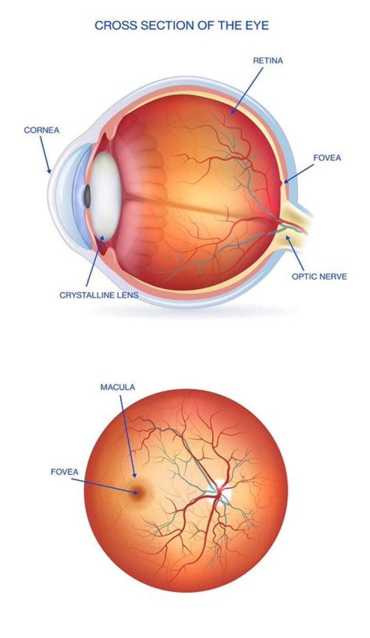
The fovea centralis, or fovea, is a small depression within the neurosensory retina where visual acuity is the highest. The fovea itself is the central portion of the macula, which is responsible for central vision.[1][2][3][4]
What is the fovea in the eye?
The depression in the very center of the macula where eyesight is sharpest. It is also called the fovea centralis. A number of eye problems can affect the fovea and can lead to vision loss if they are not treated.
Is fovea the blind spot?
The blind spot (Fovea centralis) The blind spot is located about 15 degrees on the nasal side of the fovea.
What is the main function of fovea?
The fovea is responsible for sharp central vision (also called foveal vision), which is necessary in humans for activities for which visual detail is of primary importance, such as reading and driving.What is the difference between fovea and retina?
The retina is a light-sensitive layer at the back of the eye that covers about 65 percent of its interior surface. … In the middle of the retina is a small dimple called the fovea or fovea centralis. It is the center of the eye’s sharpest vision and the location of most color perception.
How many fovea do humans have?
Total Number of Cones in Fovea approximately 200,000.
What is macula and fovea?
The macula is the pigmented part of the retina located in the very center of the retina. In the center of the macula is the fovea, perhaps the most important part of the eye. The fovea is the area of best visual acuity. It contains a large amount of cones—nerve cells that are photoreceptors with high acuity.
What does fovea look like?
The human fovea is densely packed with cones. It looks like a little pit on the retina because the cells that are above the retinal surface, such as retinal ganglion cells, horizontal cells, and amacrine cells, are swept away so that the cones are at the surface.What does the ciliary body do in the eye?
The ciliary body is found behind the iris and includes the ring-shaped muscle that changes the shape of the lens when the eye focuses. It also makes the clear fluid that fills the space between the cornea and the iris.
What happens if the fovea is damaged?When the fovea is compromised by disease or injury, the brain works, subconsciously, to find a position in the retina that it can use to develop a new fixation point — a pseudofovea — in a region of the retina with surviving photoreceptors.
Article first time published onWhat is the difference between a blind spot and the fovea?
Visual acuity such as sharpness and detail is greatest at the fovea, while at the blind spot it is insensitive to visual stimulation, it’s the part of the retina that converges to the optic nerve.
What is the difference between fovea and yellow spot?
The term “yellow spot” is another word for macula lutea or fovea: Different terms are used in different texts but the meaning is the same. Because it contains a large number of the light-sensitive photo-detector cells called cones, the yellow spot (“macula lutea”, or “fovea”) is the area of greatest acuity of vision.
Is fovea and yellow spot same?
The yellow spot or macula is an oval yellow spot near the centre of the retina of the human eye. … It is the area of best vision where maximum amount of cone cells are present.It is also known as fovea centralis and Macula Lutea. Most of the sensory cells are present at this spot.
What is macula in human eye?
The macula is part of the retina at the back of the eye. It is only about 5mm across but is responsible for our central vision, most of our colour vision and the fine detail of what we see. The macula has a very high concentration of photoreceptor cells – the cells that detect light.
What is the difference between macula lutea and fovea?
It is the part of the retina that is responsible for sharp, detailed central vision (also called visual acuity). The macula lutea, also called fovea, contains a very high concentration of cones. These are the light-sensitive cells in the retina that give detailed central vision.
Does the fovea contain cones?
The only photoreceptors located in the center of the fovea are cones. These are tightly packed, and the outer segments are elongated, appearing rodlike in shape yet containing the visual pigments of the cone population.
What is dry macular?
Dry macular degeneration is a common eye disorder among people over 50. It causes blurred or reduced central vision, due to thinning of the macula (MAK-u-luh). The macula is the part of the retina responsible for clear vision in your direct line of sight.
What is drusen made of?
Drusen are about the width of a pinhead and are composed of a mixture of proteins and lipids (naturally occurring molecules that include fats). They often cause no symptoms, but can occasionally cause visual distortion if they are very large and near the center of the retina.
What type of protein is rhodopsin?
Structurally, rhodopsin is classified as a chromoprotein (chromo is a Greek-derived root meaning “colour”). It is made up of opsin (a colourless protein) and 11-cis-retinal (11-cis-retinaldehyde), a pigmented molecule derived from vitamin A.
Are blue cones in the fovea?
Cone Details They provide the eye’s color sensitivity. The green and red cones are concentrated in the fovea centralis . The “blue” cones have the highest sensitivity and are mostly found outside the fovea, leading to some distinctions in the eye’s blue perception.
What kind of cells does the fovea have?
The fovea is not recognizable at this stage, because the central region of the retina, where the fovea will develop, consists primarily of several layers of ganglion cell bodies and inner nuclear layer cells (INL), presumably amacrine and bipolar cells (Figure 8, a).
What is the thinnest part of the retina?
The retinal thickness shows greatest variations in the center. The retina is thinnest at the foveal floor (0.10, 0.150-0.200 mm) and thickest (0.23, 0.320 mm) at the foveal rim. Beyond the fovea the retina rapidly thins until the equator.
What are ciliary muscles?
Ciliary muscle: A circular muscle that relaxes or tightens the zonules to enable the lens to change shape for focusing. The zonules are fibers that hold the lens suspended in position and enable it to change shape during accommodation.
How do you treat ciliary body?
Conservative treatment options include brachytherapy, local resection and/or cryotherapy in selected cases. We report for the first time the use of targeted chemotherapy to treat a ciliary body medulloepithelioma with aqueous and vitreous seeding.
Why is vision clearest at the fovea?
The resolution or sharpness in vision is because of the high concentration of cone cells in the fovea. The fovea has the densest concentration of photoreceptor cells that are known as cones. Rods are completely absent from the fovea. … The cones are responsible for color vision and perception of fine detail.
Why does fovea only contain cones?
In the fovea, there are NO rods… only cones. The cones are also packed closer together here in the fovea than in the rest of the retina. Also, blood vessels and nerve fibers go around the fovea so light has a direct path to the photoreceptors.
What causes fovea damage?
Conditions that may affect the fovea include: Macular degeneration (both wet and dry forms) — Age-related thinning and abnormal protein growth on the macula (dry) or macular scarring due to abnormal blood vessel growth and leakage (wet).
Can damaged retina repair itself?
Yes, in many cases an eye doctor can repair a damaged retina. While a patient may not experience completely restored vision, retinal repair can prevent further vision loss and stabilize vision. It’s important that patients get treatment for their damaged retinas as soon as possible.
What are the optic chiasm?
The place in the brain where some of the optic nerve fibers coming from one eye cross optic nerve fibers from the other eye. Also called optic chiasm.
What is the difference between myopia and Hypermetropia?
The difference between myopia and hyperopia is whether you have difficulty seeing up close or at a distance. Hyperopia (farsightedness) makes it hard to see things that are close, and Myopia (nearsightedness) makes it difficult to see things that are far away.
What are cones psychology?
The cones are receptor cells that help us see fine details of things and tend to help us see in situations where there is light or daylight. The majority of cones are in the center of the retina (we have approximately 6 million cones in each eye). … Cones also help us with color perception.