What type of stain is hematoxylin
Hematoxylin has a deep blue-purple color and stains nucleic acids by a complex, incompletely understood reaction. Eosin is pink and stains proteins nonspecifically. In a typical tissue, nuclei are stained blue, whereas the cytoplasm and extracellular matrix have varying degrees of pink staining.
What type of dye is haematoxylin?
Haematoxylin can be considered as a basic dye (general formula for basic dyes is:dye+ Cl-). Haemotoxylin is actually a dye called hematein (obtained from the log-wood tree) used in combination with aluminium ions (Al3+). It is used to stain acidic (or basophilic) structures a purplish blue.
What is hematoxylin used to stain?
Hematoxylin is used to illustrate nuclear detail in cells. Depth of coloration is not only related to the amount of DNA in the nuclei but also to the length of time the sample spends in hematoxylin.
What type of staining is hematoxylin and eosin?
H&E is the combination of two histological stains: hematoxylin and eosin. The hematoxylin stains cell nuclei a purplish blue, and eosin stains the extracellular matrix and cytoplasm pink, with other structures taking on different shades, hues, and combinations of these colors.Does hematoxylin stain acidic or basic?
Hematoxylin, a natural dye product, acts as a basic dye that stains blue or black.
Is hematoxylin a basic dye?
Haematoxylin can be considered as a basic dye. It is used to stain acidic structures a purplish blue. DNA in the nucleus, and RNA in ribosomes and in the rough endoplasmic reticulum are both acidic, and so haemotoxylin binds to them and stains them purple.
What color does hematoxylin stain structures?
Hematoxylin has a deep blue-purple color and stains nucleic acids by a complex, incompletely understood reaction. Eosin is pink and stains proteins nonspecifically. In a typical tissue, nuclei are stained blue, whereas the cytoplasm and extracellular matrix have varying degrees of pink staining.
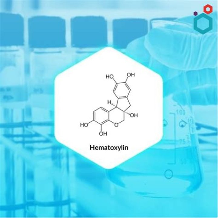
How is hematoxylin made?
Hematoxylin is a natural product extracted from the heartwood of the logwood tree (Haematoxylum campechianum).What is histological staining?
Histological staining is a series of technique processes undertaken in the preparation of sample tissues by staining using histological stains to aid in the microscope study (Anderson, 2011).
How do you make a hematoxylin stain?Method – Dissolve the hematoxylin in absolute alcohol and ammonium alum in hot water. Mix the two solutions and heat to boiling. Remove from flame, and add mercuric oxide and cool rapidly. Glacial acetic acid if added gives brisk nuclear staining, but life of the solution is reduced.
Article first time published onWhat is hematoxylin in histopathology?
Hematoxylin and eosin (H&E) is the most widely used stain in histology and allows localization of nuclei and extracellular proteins. Hematoxylin, not a dye itself, produces the blue Hematin via an oxidation reaction with nuclear histones causing nuclei to show blue.
Is hematoxylin a basophilic?
Cell structures which bind to basic dyes such as hematoxylin are said to be: basophilic.
Is hematoxylin a fluorescent?
Hematoxylin has broad absorption between 400 and 700 nm, with virtually no fluorescence emission. …
What is basic staining?
Basic stains, such as methylene blue, Gram safranin, or Gram crystal violet are useful for staining most bacteria. These stains will readily give up a hydroxide ion or accept a hydrogen ion, which leaves the stain positively charged.
What does trichrome stain?
Trichrome staining is used to visualize connective tissues, particularly collagen, in tissue sections. In a standard Masson’s Trichrome procedure, collagen is stained blue, nuclei are stained dark brown, muscle tissue is stained red, and cytoplasm is stained pink.
Is hematoxylin positive or negative?
Haematoxylin in complex with aluminium salts is cationic and acts as a basic dye. It is positively charged and can react with negatively charged, basophilic cell components, such as nucleic acids in the nucleus. These stain blue as a result.
What is hematoxylin and eosin staining used for?
H and E staining helps identify different types of cells and tissues and provides important information about the pattern, shape, and structure of cells in a tissue sample. It is used to help diagnose diseases, such as cancer. Also called hematoxylin and eosin staining.
What is the difference between hematoxylin and eosin?
Hematoxylin and eosin are important dye compounds in staining microstructures such as proteins in the cytoplasm. The key difference between hematoxylin and eosin is that hematoxylin is a basic dye, whereas eosin is an acidic dye.
What is regressive hematoxylin?
In the regressive H&E staining method, hematoxylin is applied to the tissue section and is then followed by a differentiating solution specifically designed to remove excess stain from the nuclei. Strong or weak acids may be used as components of the differentiating solution.
What is the pH of hematoxylin?
The pH and peak of absorbance of the aliquots were pH = 2.0 450 NM, 2.5 505, 2.6 507, 2.7 515, 2.8 520, 2.9 530, 3.0 540, 3.1 550, 3.3 560, 3.5 560. In the stained material in the intensity of nuclear staining was about the same at all pH levels but non-specific staining was greatest in slides stained at pH = .
What is the active coloring agent of hematoxylin?
The active ingredient in hematoxylin solutions is hematein complexed with a metal ion-eg, aluminum, iron, tungsten. Aluminum is the most commonly used. If aluminum is used, the hematoxylin solution will stain blue; if iron is used hematoxylin will stain black or blue-black.
What color do basophils stain?
Basophils are the least numerous of the granulocytes and account for less than 1 percent of all white blood cells occurring in the human body. Their large granules stain purple-black in colour and almost completely obscure the underlying double-lobed nucleus.
What are the different types of staining?
- Staining Type # 2. Differential Staining:
- Staining Type # 3. Gram Staining:
- Staining Type # 4. Acid Fast Staining:
- Staining Type # 5. Endospore Staining:
What is histochemical staining?
Histochemistry involves the differential staining of cells (i.e., using dyes that stain specific structural and molecular components) to reflect the chemical differences of the constituents.
Which stain is used in the demonstration of carbohydrates?
Periodic Acid Schiff (PAS) Staining: A Useful Technique for Demonstration of Carbohydrates.
What is the active ingredient of hematoxylin?
Although the stain is commonly called haematoxylin, the active colourant is the oxidized form haematein, which forms strongly coloured complexes with certain metal ions (commonly Fe(III) and Al(III) salts).
How do you dilute hematoxylin?
I often dilute a city water (1:10 dilution). Then kept in the dark at least one night to remove some precipitate. Carefully collect the diluted solution without mixing the bottom. The diluted solution never overstain.
How do you remove hematoxylin stains?
1% Acid Alcohol should do the trick just fine. HCL in 70% ethanol. Rinse either with 0.25% hydrochloric acid (HCl) for 2-5 seconds or 1% acid alcohol (1ml Conc HCl in 100ml ethanol) to remove excess stain from the slide, Then keep the slides in running water for 3 minutes for blueing.
What is a basophilic stain?
Basophilic describes the appearance of structures seen in histological sections which take up basic dyes. The structures usually stained are those that contain negative charges, such as the phosphate backbone of DNA in the cell nucleus and ribosomes.
Is the nucleus acidophilic or basophilic?
Nuclei are basophilic and are stained blue. At lower magnifications they appear as blue dots and at higher magnifications chromatin and nucleoli may be identified within the nucleus. Surrounding the nucleus is the acidophilic cytoplasm stained pink (due to the positive charges on arginine and lysine).
Are ribosomes eosinophilic or basophilic?
Most cellular organelles and extracellular matrix are eosinophilic, while the nucleus, rough endoplasmic reticulum, and ribosomes are basophilic.