Where are the Leptomeninges
The leptomeninges, or the pia and arachnoid mater
Where are the meninges located and what is their function?
The meninges is a layered unit of membranous connective tissue that covers the brain and spinal cord. These coverings encase central nervous system structures so that they are not in direct contact with the bones of the spinal column or skull.
Where is the subarachnoid located?
It is located at the lower lumbar spinal canal. It extends from the conus medullaris around the level of the first and second lumbar vertebrae to the level of the second sacral vertebra. It contains the filum terminale and the cauda equina.
Where are the meninges located in the brain?
Brain meninges are three-layer tissue envelopes that have a protective, supportive and metabolic role. They are located between the brain and the skull and between the spinal cord and spinal vertebrae and are constructed of loose and dense connective tissues.What is the meaning of Leptomeninges?
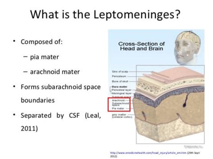
Leptomeninges: The two innermost layers of tissue that cover the brain and spinal cord. The two layers are called the arachnoid mater and pia mater.
Which organ is surrounded by meninges?
meninges, singular meninx, three membranous envelopes—pia mater, arachnoid, and dura mater—that surround the brain and spinal cord. Cerebrospinal fluid fills the ventricles of the brain and the space between the pia mater and the arachnoid.
Where is the origin of the meninges?
The cranial meninges originate from a mesenchymal sheath on the surface of the developing brain, called primary meninx, and undergo differentiation into three layers with distinct histological characteristics: the dura mater, the arachnoid mater, and the pia mater.
What part of the nervous system is the brain?
The brain and the spinal cord are the central nervous system. The nerves that go through the whole body make up the peripheral nervous system.Where do meninges end?
While it has two layers in the cranial segment, the spinal dura mater only has the deep meningeal layer. The periosteal layer, which is the superficial layer of the dura within the calvarium, ends at the foramen magnum, with only the meningeal layer continuing down along the spinal cord.
Is the subarachnoid space outside the brain?Cerebrospinal fluid circulates within the ventricles of the brain and in the subarachnoid space outside the brain.
Article first time published onHow does subarachnoid space protect the brain?
The subarachnoid space is the cerebrospinal fluid-filled space that exists between the arachnoid and the pia. … The primary functions of the CSF are to cushion the brain and spinal cord from trauma and to supply them with nutrients and remove waste.
How does blood get into the subarachnoid space?
Most often, it occurs when a weak area in a blood vessel (aneurysm) on the surface of the brain bursts and leaks. The blood then builds up around the brain and inside the skull increasing pressure on the brain. This can cause brain cell damage, life-long complications, and disabilities.
How long can you live with leptomeningeal carcinomatosis?
Prognosis remains grim in patients with LC. The time from diagnosis to death is about 4 to 6 weeks if left untreated. With treatment, overall survival is approximately 2 to 4 months.
What is the role of the meninges?
Three layers of membranes known as meninges protect the brain and spinal cord. The delicate inner layer is the pia mater. The middle layer is the arachnoid, a web-like structure filled with fluid that cushions the brain.
What are the symptoms of leptomeningeal carcinomatosis?
- Headaches.
- Nausea (feeling like you’re going to throw up) or vomiting (throwing up)
- Difficulty thinking.
- Double vision.
- Dizziness.
- Difficulty speaking or swallowing.
- Pain in your arms and legs.
- Weakness or lack of coordination in your arms and legs.
Where does pia mater end?
In the brain, this ends up in the interstitial space. The protein portions are able to leave through the very permeable pia mater and enter the subarachnoid space in order to flow in the cerebrospinal fluid (CSF), eventually ending up in the cerebral veins.
What is underneath the arachnoid layer?
The space under the arachnoid, the subarachnoid space, is filled with cerebrospinal fluid and contains blood vessels. The pia mater is the innermost layer of meninges. This thin, delicate membrane is tightly bound to the surface of the brain and spinal cord and cannot be dissected away without damaging the surface.
Where does the arachnoid mater end?
The arachnoid mater and dura mater are very close together throughout the cranium and spinal canal all the way to S2, where the two layers fuse into one and end in the filum terminale, which attaches to the coccygeal end of the spinal canal.
What is meninges in biology?
The meninges are membranous layers surrounding the central nervous system. … For the brain, the meninges regulate diverse processes including cell survival, cell migration, generation of neurons from progenitors, and vascularization. Also, the meninges serve as a stem cell niche for the brain in the postnatal life.
Which organ of the body is surrounded by?
brain and spinal cord of vertebrates are the body organs which are surrounded by meninges.
What are the meninges quizlet?
Protect the brain from injury and is made up of three layers: dura mater, arachnoid mater, and pia mater. Two layers: superficial Periosteal Layer attachs to the skull and deeper Meningeal Layer forms the true external covering of the brain.
What is the epidural space?
The epidural space is the area between the dura mater (a membrane) and the vertebral wall, containing fat and small blood vessels. The space is located just outside the dural sac which surrounds the nerve roots and is filled with cerebrospinal fluid.
Where is the CSF located in the spinal meninges?
The CSF is contained within the subarachnoid space, between the arachnoid mater and the pia mater layers.
Where do somatic motor neurons reside?
Somatic motor neurons. Somatic MNs are located in the Rexed lamina IX in the brainstem and the spinal cord and innervate skeletal muscles responsible for movements (Rexed, 1954). MNs form coherent groups connecting to a unique muscle target defined as MN pools.
What are the 5 main parts of the nervous system?
- Afferent, Efferent, and Mixed Nerves. …
- Cranial Nerves. …
- Spinal Nerves.
What are the 3 types of the brain?
The brain can be divided into three basic units: the forebrain, the midbrain, and the hindbrain. The hindbrain includes the upper part of the spinal cord, the brain stem, and a wrinkled ball of tissue called the cerebellum (1).
What are the 3 nervous systems?
It has three parts: The sympathetic nervous system. The parasympathetic nervous system. The enteric nervous system.
What is the largest cistern?
Cisterna magna also called cerebellomedullary cistern – the largest of the subarachnoid cisterns. It lies between the cerebellum and the medulla oblongata. It receives CSF from the fourth ventricle via the median aperture (foramen of Magendie).
Where is the subdural space?
The subdural space is a potential intracranial space situated between the arachnoid and dura. Fluid can collect in the subdural space and in the subarachnoid space.
Does the central canal contain CSF?
The central canal is part of a system of cerebrospinal fluid (CSF) cavities that includes the cerebral ventricle, aqueduct of Sylvius, and fourth ventricle (Figures 3-4) [2]. It is situated in the gray commissure, which (along with the anterior white commissure) connects the two parts of the spinal cord.
Which bones protect the brain?
The skull protects the brain and forms the shape of the face. The spinal cord, a pathway for messages between the brain and the body, is protected by the backbone, or spinal column.